Procedures
Advanced Implant Solutions
For select patients with severe upper‑jaw bone loss, remote anchorage options—such as zygomatic (cheekbone) implants and pterygoid implants—may provide stable support when conventional implants are limited.
When bone loss is severe
The upper jaw (maxilla) can lose bone after long‑standing tooth loss, periodontal disease, trauma, or prior extractions. In some cases, the maxillary sinuses expand as bone resorbs, leaving limited height and width for standard implant placement.
What “remote anchorage” means
Remote anchorage techniques use stronger, more stable bone away from the resorbed ridge to support an implant‑retained restoration. This approach can reduce the need for extensive bone grafting in select cases, and it can be especially helpful for patients considering full‑arch implant surgery (All‑on‑X).
Download: All‑on‑X Recovery Guide (PDF)
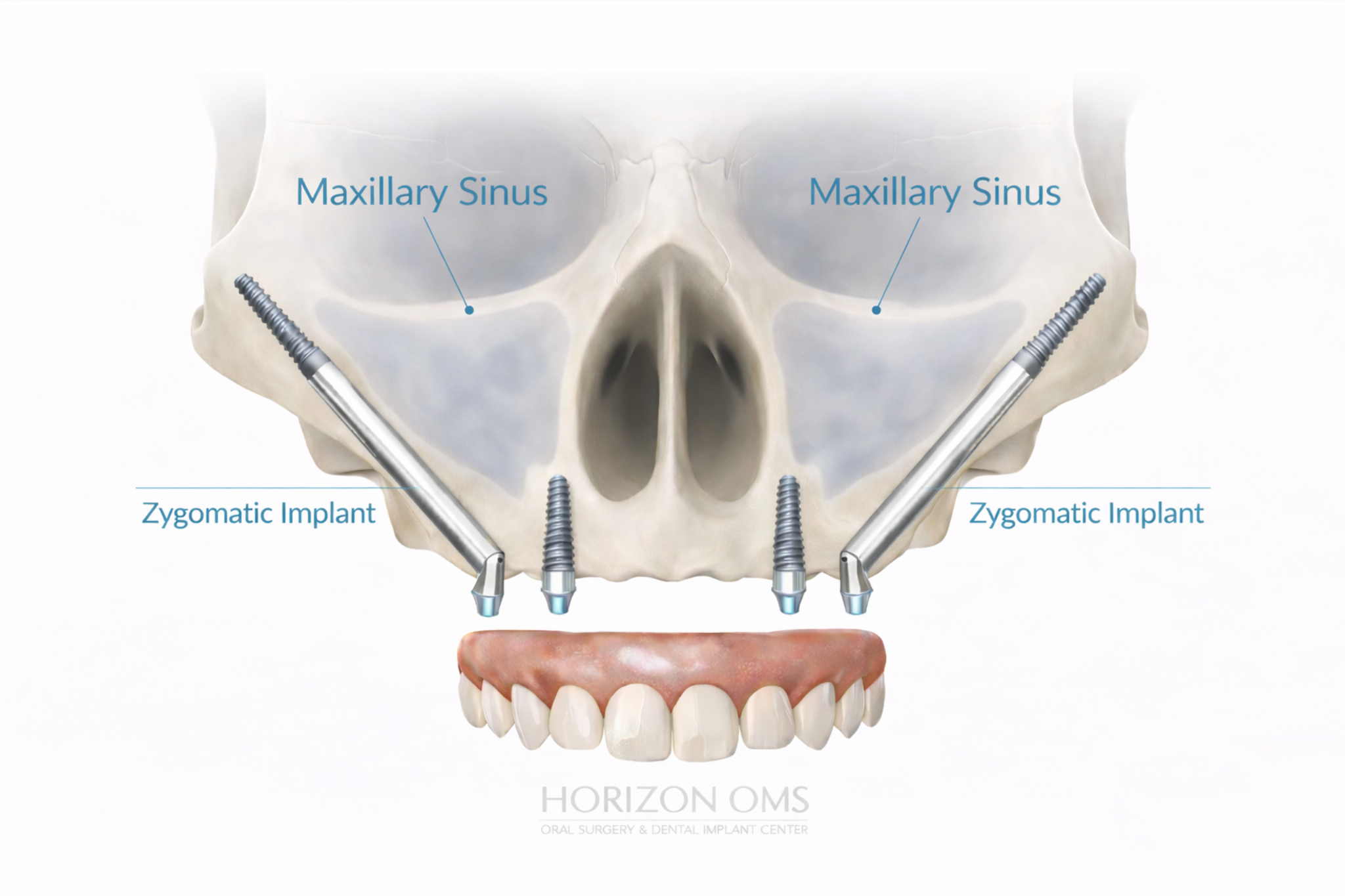
Zygomatic implants (cheekbone implants)
Zygomatic implants are longer implants that gain stability in the zygoma (cheekbone). They are used in the upper jaw when the back portion of the maxilla has lost significant bone. Because this region sits near important structures—such as the maxillary sinus and the area around the orbit (eye socket)—planning requires detailed knowledge of facial anatomy and careful 3D imaging.
Clinical reference: Example panoramic X‑ray showing zygomatic implant placement in a severely resorbed upper jaw.
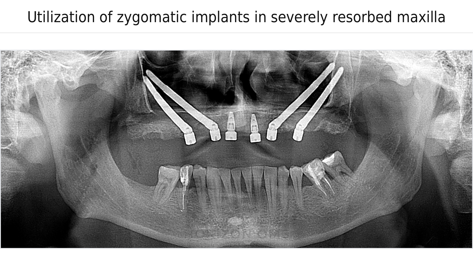
Panoramic radiograph shown for educational reference. Final recommendations require CBCT imaging and prosthetic‑driven planning.
Pterygoid implants
Pterygoid implants gain anchorage in dense bone behind the upper jaw near the pterygoid plates. They can provide posterior (back‑of‑the‑jaw) support when bone is limited near the sinus region. In select cases, they can complement a full‑arch plan and reduce the need for sinus grafting.
How we plan safely
- CBCT (3D imaging) to evaluate bone, sinus anatomy, and key landmarks
- Prosthetic‑driven planning in coordination with your restoring dentist to confirm the final tooth position first
- Digital planning to determine implant number, angulation, and anchorage sites
- Anesthesia options tailored to the procedure and your health history, including office‑based IV sedation when appropriate
How this fits with your overall implant plan
Many patients exploring advanced implant solutions are also considering traditional options such as standard dental implants and bone grafting. During your consultation, we’ll review imaging, explain the pros/cons of each approach, and coordinate with your restorative team to choose the most predictable plan.